Our Research
-
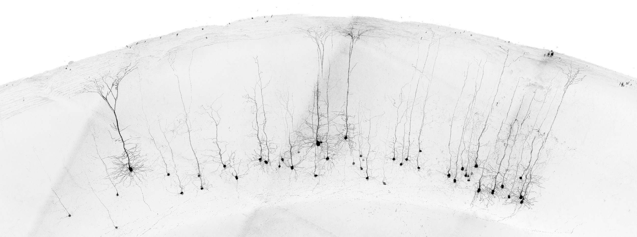
How are memories made?
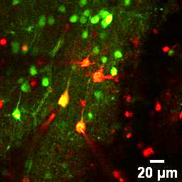
Understanding how information is encoded, stored in, and retrieved from memory by mammalian brain circuits is a central drive of the lab. We use multi-photon calcium imaging to study the activity of populations of neurons in the hippocampus (and connected areas) of mice while they are performing simple cognitive tasks. We are investigating episodic memory, working memory and spatial memory.
-
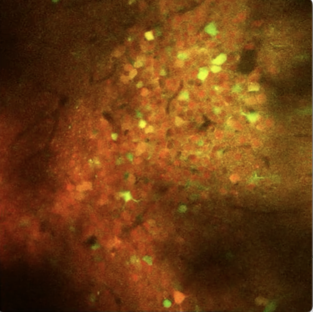
Alzheimer's Disease
We are interested in how information processing for memory in hippocampal and cortical circuits is affected by the pathophysiology of Alzheimer’s Disease, and how it can be restored following therapeutic treatment. We have developed a high content assay which allows us to track alterations in circuit function, behavioural performance and 3D distribution of gene expression and/or amyloid aggregation throughout the whole brain, in the same mice.
-
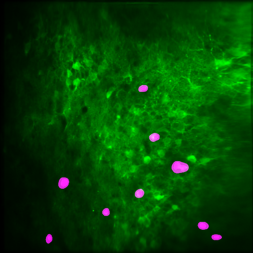
Cerebral organoids
Animal models of neurodegenerative diseases have many advantages, but suffer from the issue that they are not based on human genetics. We have recently begun a project using 3D cerebral organoid cultures based on human stem cells to characterise and treat neurodegenerative disease processes. We use a viral approach to genetically modify the organoids, and use two-photon imaging to observe progressive changes in organoid spatiotemporal neural activity dynamics.
-
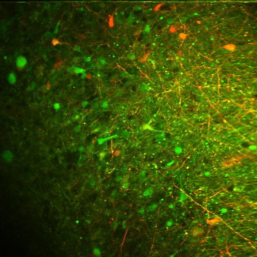
Neurotechnology
We develop novel technology for interrogating and modulating brain circuits, with a particular focus on the use of light. Recent advances have included two-photon targeted robotic patch-clamp electrophysiology (the picture here shows a robotic quad patch-clamp recording) and source-localised multifocal multiphoton microscopy.
-
Neurophysiological data analysis
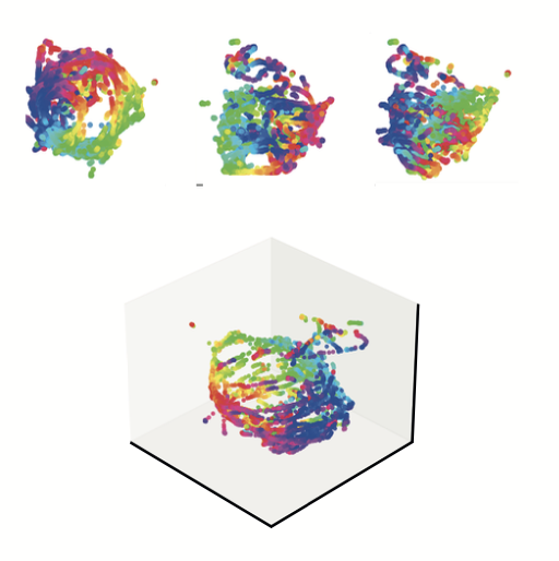
New technology allows us to record from hundreds or thousands of neurons simultaneously, across widespread brain areas. Interpreting this data requires new theoretical approaches. We develop and use tools based on information theory, network theory and the physics of complex systems to analyse large-scale neural datasets.
-

Facilities
We have several two-photon microscopes suitable for both in vitro and in vivo mouse brain imaging, a three-photon microscope for in vivo deep tissue imaging, and a Tissuecyte-1000 ex vivo whole organ two-photon scanning microtome. We are currently building a two-photon mesoscope for imaging large fields of view (5mm) or multiple regions simultaneously. We also have access to cerebral organoid cell culture facilities.
We are grateful to have received funding for our research in recent years from Wellcome, BBSRC, EPSRC, and from Mrs Anne Uren and the Michael Uren Foundation.